
Diabetes & Heart Failure Risk Calculator
People with diabetes have significantly higher rates of:
- Hypertension: 68% vs 32%
- Obesity (BMI ≥ 30): 57% vs 23%
- Elevated LDL-C: 61% vs 27%
Your Risk Assessment
Did you know that adults with diabetes are up to twotimes more likely to end up with left ventricular failure than those without? The link isn’t a coincidence-high blood sugar, insulin resistance, and associated conditions like hypertension silently reshuffle the heart’s pumping power. This article breaks down the biology, the warning signs, and what you can do today to keep your heart beating strong.
Key Takeaways
- Persistent high glucose and insulin resistance damage heart muscle cells, leading to reduced ejection fraction.
- Hypertension and obesity, common in diabetes, double the risk of left ventricular failure.
- Early detection through echocardiography and biomarkers can catch the problem before symptoms appear.
- Managing blood sugar, blood pressure, and weight together offers the best chance to prevent or slow heart decline.
- Targeted medications-ACE inhibitors, SGLT2 inhibitors, and beta‑blockers-address both diabetes and heart stress.
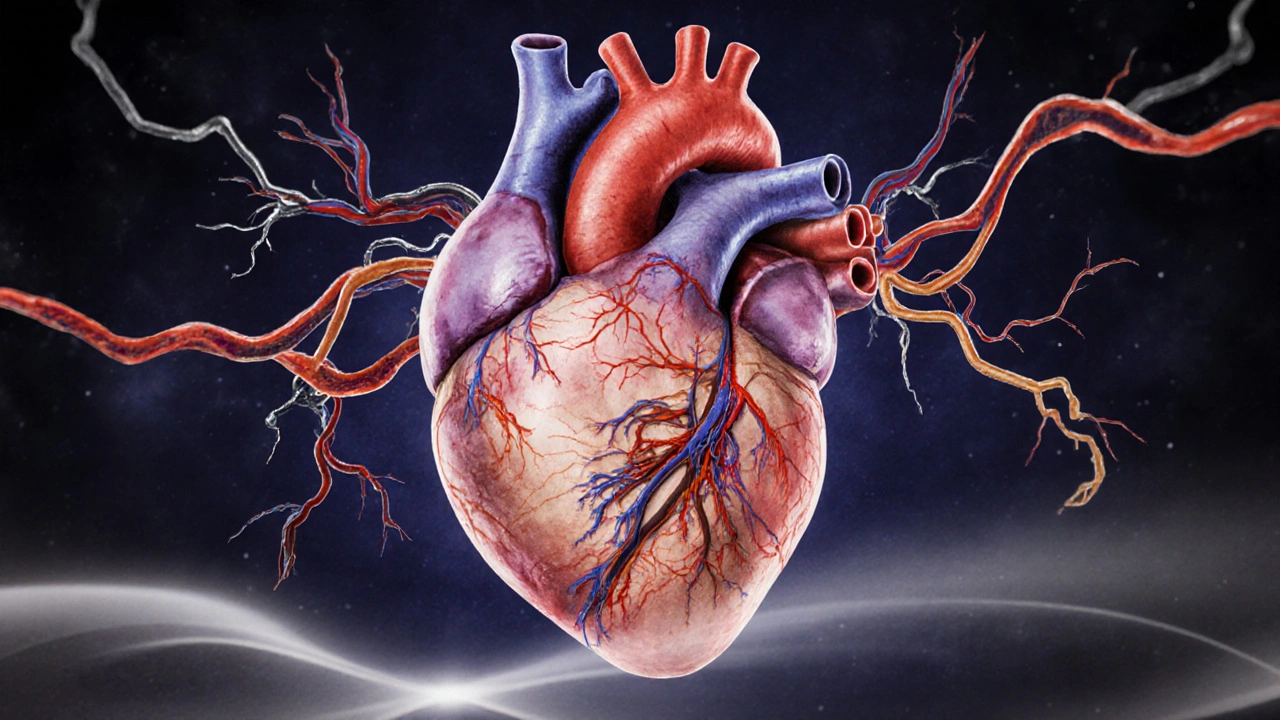
Understanding Left Ventricular Failure
Left Ventricular Failure a condition where the left pumping chamber of the heart can’t push enough blood to meet the body’s needs is the most common form of heart failure. When the left ventricle stiffens or weakens, blood backs up into the lungs, causing shortness of breath, fatigue, and fluid buildup. The hallmark measurement is a low Ejection Fraction the percentage of blood pumped out of the left ventricle with each beat, typically below 40% in symptomatic patients.
Why Diabetes Is a Major Driver
Diabetes a chronic metabolic disease characterized by elevated blood glucose levels affects the heart through several intertwined pathways:
- Hyperglycemia‑induced toxicity: Excess glucose creates advanced glycation end‑products (AGEs) that stiffen cardiac tissue and impair relaxation.
- Insulin resistance: When cells stop responding to insulin, the heart shifts to fatty‑acid metabolism, which is less efficient and generates harmful oxidative stress.
- Microvascular disease: Small‑vessel damage reduces oxygen delivery to heart muscle, promoting Cardiomyopathy a disease of the heart muscle that weakens its ability to contract.
- Inflammation: Chronic low‑grade inflammation raises cytokine levels, which blunt contractility and promote fibrosis.
- Co‑existing hypertension and obesity: Both are more prevalent in diabetic populations and further strain the left ventricle.
In short, diabetes creates a perfect storm that chips away at the heart’s pumping capacity.
Risk Factors at a Glance
| Risk Factor | Diabetes (%) | Non‑Diabetes (%) |
|---|---|---|
| Hypertension | 68 | 32 |
| Obesity (BMI≥30) | 57 | 23 |
| Elevated LDL‑C | 61 | 27 |
| Reduced Ejection Fraction (<40%) | 15 | 6 |
| Microvascular Complications | 42 | 9 |
Spotting the Early Signs
Because left ventricular failure can develop quietly, clinicians rely on objective tests:
- Echocardiography: Measures ejection fraction and detects wall thickening.
- Biomarkers: Elevated B‑type natriuretic peptide (BNP) or NT‑proBNP signals cardiac stress.
- Electrocardiogram (ECG): May reveal prior silent myocardial infarctions common in diabetics.
Patients often report fatigue after mild activity, unexplained weight gain from fluid retention, or waking up short‑of‑breath. If you have diabetes and notice any of these, it’s time to ask your doctor for a cardiac screen.
Managing Diabetes to Protect the Left Ventricle
Effective management tackles three fronts: blood sugar, blood pressure, and body weight.
- Glycemic control: Aim for an A1C below 7% (or individualized target). Newer agents like SGLT2 inhibitors not only lower glucose but also reduce heart‑failure hospitalizations.
- Blood pressure: Keep systolic pressure under 130mmHg. ACE inhibitors and ARBs are first‑line because they relax the vessels and lessen ventricular remodeling.
- Weight reduction: A 5‑10% weight loss can improve insulin sensitivity and lower left‑ventricular wall stress.
Exercise remains a cornerstone. Moderate‑intensity aerobic activity-30minutes, five days a week-enhances endothelial function and aids glucose uptake without over‑taxing the heart.

Pharmacologic Tools That Target Both Conditions
Several drug classes hit two birds with one stone:
- SGLT2 inhibitors (e.g., empagliflozin): Cut glucose reabsorption in kidneys and have a proven benefit in reducing heart‑failure events.
- GLP‑1 receptor agonists: Promote weight loss, improve blood pressure, and may modestly boost cardiac output.
- Beta‑blockers: Slow heart rate, lower oxygen demand, and blunt the adverse effects of sympathetic overdrive common in diabetics.
Combination therapy should be personalized; discuss potential side effects like dehydration (with SGLT2 inhibitors) or bradycardia (with beta‑blockers) with your clinician.
Preventive Checklist for Diabetic Patients
- Schedule an echocardiogram at diagnosis if you have type2 diabetes over age40.
- Check BNP or NT‑proBNP annually if you have hypertension or a history of myocardial infarction.
- Maintain A1C within target range; adjust therapy promptly after any flare‑up.
- Control blood pressure with regular home monitoring; aim for < 130/80mmHg.
- Adopt a Mediterranean‑style diet rich in leafy greens, fatty fish, and whole grains.
- Engage in at least 150minutes of moderate‑intensity activity per week.
- Quit smoking; each pack‑year adds about a 10% risk for heart‑failure hospitalization.
Following these steps can delay or even prevent the onset of left ventricular failure, keeping you active and independent.
Frequently Asked Questions
Can type1 diabetes also cause left ventricular failure?
Yes. Although type2 diabetes is more prevalent, chronic hyperglycemia in type1 patients still drives myocardial fibrosis and microvascular damage, raising the heart‑failure risk.
Is an echocardiogram necessary for every diabetic?
Not mandatory for all, but guidelines recommend a baseline echo for diabetics over 40 or those with additional risk factors like hypertension, obesity, or known coronary disease.
Do SGLT2 inhibitors replace heart‑failure medicines?
No. They complement standard heart‑failure drugs (ACE inhibitors, beta‑blockers) and have been shown to reduce hospital admissions when added to the regimen.
What lifestyle change has the biggest impact?
Weight loss combined with regular aerobic exercise provides the strongest dual benefit-improving insulin sensitivity and lowering ventricular after‑load.
How quickly can left ventricular function improve after better glucose control?
Improvements are modest and often take 6-12months. Early intervention yields the best chance of reversing subtle dysfunction before permanent fibrosis sets in.


20 Comments
Diabetes really does double the risk of left‑ventricular failure, so taking care of blood sugar is essential.
It’s fascinating how metabolic pathways intersect with cardiac mechanics; the cascade of insulin resistance, inflammation, and fibrotic remodeling paints a complex picture :)
Just to add, the article missed the fact that advanced glycation end‑products (AGEs) also affect the extracellular matrix – that’s why you see stiffening of the ventricle in many diabetics. Also, microvascular disease isnt just a foot problem, it hits the heart too.
Great summary keep it up love the clear breakdown of risk factors and practical tips
When we talk about left ventricular failure in the context of diabetes we have to first acknowledge the sheer burden of hyperglycemia on myocardial cells. The chronic exposure to elevated glucose concentrations leads to the formation of advanced glycation end‑products, which in turn cross‑link collagen fibers and reduce compliance. On top of that, insulin resistance forces the heart to rely more heavily on fatty‑acid oxidation, a less efficient energy source that generates reactive oxygen species. Those oxidative stressors damage mitochondrial DNA and further impair contractile function. The microvascular network isn’t spared either; capillary rarefaction diminishes oxygen delivery, creating ischemic patches in the myocardium. Inflammation follows, as cytokines like TNF‑α and IL‑6 promote fibroblast activation and extracellular matrix deposition. The resulting fibrosis stiffens the ventricular wall, raising filling pressures and precipitating diastolic dysfunction. Hypertension, which is prevalent in up to 68 % of diabetic patients, adds afterload stress that the compromised left ventricle struggles to overcome. Obesity compounds the issue by increasing circulating volume and raising systemic vascular resistance. Moreover, dyslipidemia, particularly elevated LDL‑C, accelerates atherosclerotic plaque formation, narrowing coronary arteries and limiting perfusion. All these mechanisms converge to erode ejection fraction over time, often silently until symptoms emerge. Early detection through echocardiography can reveal subtle reductions in strain before a drop in EF becomes apparent. Biomarkers such as NT‑proBNP also rise in response to ventricular wall stress, offering a laboratory clue. Therapeutically, SGLT2 inhibitors have shown promise by reducing cardiac preload and improving metabolic efficiency. ACE inhibitors and ARBs temper the renin‑angiotensin‑aldosterone system, lowering afterload and slowing remodeling. Beta‑blockers help control heart rate, giving the ventricle more time to fill. Lifestyle interventions-tight glycemic control, regular aerobic exercise, weight loss, and low‑salt diets-remain foundational. In essence, diabetes creates a perfect storm of metabolic, hemodynamic, and inflammatory insults that collectively undermine left ventricular performance, making a comprehensive, multidisciplinary approach indispensable for prevention and management.
Oh, the tragedy of a heart that’s been silently battered by sugar! Each glucose molecule feels like a tiny dagger, chipping away at the muscle’s resilience, and before you know it, the left ventricle is gasping for breath like a child learning to swim in a storm. The article captures the clinical facts, but the emotional weight of watching loved ones struggle with fatigue and shortness of breath is something no chart can convey. It’s a relentless echo of hope and fear, where the promise of a new medication feels like a lifeline, yet the everyday battle with diet and exercise becomes an endless maze. I’m reminded of how the body’s language is subtle; a slight swelling of ankles, a whisper of breathlessness at night-these are the heart’s cries for help. So let’s not just read the numbers, let’s feel the urgency and push forward with every ounce of compassion we can muster.
From a clinical perspective, the interplay of hyperglycemic toxicity and neurohormonal activation is a textbook example of pathophysiological synergy. The elevated AGEs not only stiffen myocardial tissue but also serve as ligands for RAGE receptors, perpetuating inflammatory cascades. When you add SGLT2 inhibitor‑mediated natriuresis into the mix, you effectively reduce preload, which can translate into measurable improvements in EF. All this underscores the importance of a multimodal therapeutic regimen that addresses both metabolic and hemodynamic axes.
Sure the article is fine but it ignores how Indian diets are the real problem the West gets to brag about healthy eating
Oh absolutely, because managing diabetes is just a walk in the park, right? I mean, who needs actual medical guidance when we have the internet?
I wonder how many patients actually get regular echocardiograms to catch early changes before symptoms appear.
Not all that useful info seems like a waste of time
Honestly the link between diabetes and heart failure is overrated; many other factors play bigger roles
Wow, another groundbreaking revelation that we’ve known for decades-thanks for the thrilling insight!
Love how this breaks it down! 🫀💪 Keep spreading the knowledge! 😊
Great read, very helpful! 👍📚
Indeed, the interrelationship between hyperglycemia, oxidative stress, and extracellular matrix remodeling is, without doubt, a multifaceted process, and, consequently, a comprehensive therapeutic strategy is paramount; moreover, the inclusion of SGLT2 inhibitors has, in recent trials, demonstrated statistically significant reductions in cardiovascular mortality, thereby affirming their role in contemporary management.
Let’s stay active, keep the sugar low, and give the heart a fighting chance!
Good summary; watch your blood pressure.
Thanks for the clear steps! I’ll start with checking my BGlc daily and schedule an echo soon. Oops I typed BGlc wrong but you get the idea. Also, counting steps and cutting soda should help too.
Oh great, another article telling us to eat salad-so original.